NEW YORK (AP) — Brain scans offer a tantalizing glimpse into the mind’s mysteries, promising an almost X-ray-like vision into how we feel pain, interpret faces and wiggle fingers.
Studies of brain images have suggested that Republicans and Democrats have visibly different thinking, that overweight adults have stronger responses to pictures of food and that it’s possible to predict a sober person’s likelihood of relapse.
But such buzzy findings are coming under growing scrutiny as scientists grapple with the fact that some brain scan research doesn’t seem to hold up.
Such studies have been criticized for relying on too few subjects and for incorrectly analyzing or interpreting data. Researchers have also realized a person’s brain scan results can differ from day to day _ even under identical conditions _ casting a doubt on how to document consistent patterns.
With so many questions being raised, some researchers are acknowledging the scans’ limitations and working to overcome them or simply turning to other tests.
Earlier this year, Duke University researcher Annchen Knodt’s lab published the latest paper challenging the reliability of common brain scan projects, based on about 60 studies of the past decade including her own.
“We found this poor result across the board,” Knodt said. “We’re basically discrediting much of the work we’ve done.”
Watching Brains `Light Up’
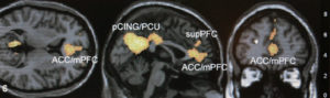
The research being re-examined relies on a technique called functional magnetic resonance imaging, or fMRI.
Using large magnets, the scans detect where oxygenated blood rushes to when someone does an activity _ such as memorizing a list of words or touching fingertips together _ allowing scientists to indirectly measure brain activity.
When the technology debuted in the early 1990s, it opened a seemingly revolutionary window into the human brain.
Other previous imaging techniques tracked brain activity through electrodes placed on the skull or radioactive tracers injected into the bloodstream. In comparison, fMRI seemed like a fast, high-resolution and non-invasive alternative.
A flurry of papers and press coverage followed the technique’s invention, pointing to parts of the brain that “light up” when we fall in love, feel pain, gamble or make difficult decisions. But as years passed, troubling evidence began to surface that challenged some of those findings.
“It’s a very powerful thing to show a picture of the brain. It lends itself to abuse, in some ways,” said Damian Stanley, a brain scientist at Adelphi University. “People eat them up, things get overblown. Somewhere in there, we lost the nuance.”
Questions Emerge
In 2009, a group of scientists investigated papers that had linked individual differences in brain activity to various personality types. They found many used a type of analysis that reported only the strongest correlations, leading to potentially coincidental conclusions. A “disturbingly large” amount of fMRI research on emotion and personality relied on these “seriously defective research methods,” the group wrote.
Later that year, another pair of researchers demonstrated that the raw results of imaging scans — without the proper statistical corrections — could detect brain activity in a dead Atlantic salmon. Four years ago, another group of scientists claimed a different common statistical error had led thousands of fMRI projects astray.
This year, Stanford University researchers described what happened when they gave the same fMRI data to 70 groups of independent neuroscientists. No two teams used the same analysis methods and, overall, the researchers did not always come to the same conclusions about what the data demonstrated about brain activity.
“In the end, we probably jumped on the fMRI bandwagon a little too fast. It’s reached the threshold of concern for a lot of us,” said Duke neuroscientist Anita Disney.
The Next Big Thing
With doubts growing, many labs have become more cautious about what imaging techniques to use in efforts to unravel the average brain’s 110,000 miles (177,000 kilometers) of nerve fibers.
Yale University researcher Joy Hirsch, for example, wants to understand “the social brain” — what happens when people talk, touch or make eye contact. She’s opted out of fMRI, since it can only be used on a single person who must remain perfectly still for imagining inside a large scanner.
Instead, Hirsch uses an alternative technology that bounces laser lights off of a fiber optic cable-laced skullcap into the brain to detect blood flow. The technique, functional near infrared spectroscopy, allows her subjects to move freely during scanning and permits her to study live social interactions between several people.
Disney also shies away from fMRI, which she says is too crude of an instrument for her forays into the molecular relationship between brain chemistry, behavior and states like arousal and attentiveness.
That doesn’t mean everyone is walking away from fMRI.
Some surgeons depend on the technique to map a patient’s brain before surgeries, and the technology has proven itself useful for broadly mapping the neural mechanisms of diseases such as schizophrenia or Alzheimer’s.
Today, optogenetics — an emerging technique that uses light to activate neurons — is poised to be brain science’s next siren technology.
Some say it’s too early to know whether they’ll adopt it as a tool.
“In that early hyper-sexy phase of a new technique, it is actually really difficult to get people to do the basic work of understanding its limitations,” Disney said.
The evolving understanding of fMRI and its limits shows science at work and should ultimately make people more confident in the results, not less, said Stanford brain scientist Russ Poldrack.
“We want to show people you have to pay attention to this stuff,” Poldrack said. “Otherwise people are going to lose faith in our ability to answer questions.”
Was this article valuable?
Here are more articles you may enjoy.
 Snap Nears Settlement of Addiction Case Ahead of Jury Trial
  After Losing Job and Crypto, Man Falsely Claimed $1.3M From 107 Class Actions
  Allianz Unit to Cut as Many as 1,800 Jobs in Push to Adopt AI
  Lawsuit Accuses Meta of Using AI to Target Workers With Medical Conditions for Layoffs
Want to stay up to date?
Get the latest insurance news
sent straight to your inbox.